Tissues of the Immune System:
The tissues of the immune system consist of the generative lymphoid organs, in which T and B lymphocytes mature and become competent to respond to antigens. And the peripheral lymphoid organs, in which adaptive immune response to microbes are initiated.
Primary Lymphoid Organs:
Bone Marrow
The bone marrow is a primary lymphoid organ that supports self-renewal and differentiation of hematopoietic stem cells (HSCs) into mature blood cells. Although all bones contain marrow, the long bones (femur, humerus), hip bones (ileum), and sternum tend to be the most active sites of hematopoiesis. The bone marrow is not only responsible for the development and replenishment of blood cells, but it is also responsible for maintaining the pool of HSCs throughout the life of an adult vertebrate.
Thymus:
T-cell precursors, which still retain the ability to give rise to multiple hematopoietic cell types, travel via the blood from the bone marrow to the thymus. Immature T cells, known as thymocytes (thymus cells) because of their site of maturation, pass through defined developmental stages in specific thymic microenvironments as they mature into functional T cells. The thymus is a specialized environment where immature T cells generate unique antigen receptors (T cell receptors, or TCRs) and are then selected based on their reactivity to self MHC-peptide complexes expressed on the surface of thymic stromal cells.
Peripheral Lymphoid Organs:
The peripheral lymphoid organs consist of Lymph nodes, Spleen and Mucosal and cutaneous immune systems. They are organized in a way that promotes the development of adaptive immune responses.
Lymph Nodes:
Lymph nodes are encapsulated nodular aggregates of lymphoid tissues located along lymphatic channels throughout the body. As the lymph passes through lymph nodes, APCs in the nodes can sample the antigens of microbes that may enter through epithelia into tissues. Dendritic cells pick up antigens of microbes from epithelia and other tissues and transport these antigens to the lymph nodes.
Spleen:
The spleen is a highly vascularized abdominal organ that serves the same role in immune responses to blood-borne antigens as that of lymph nodes in responses to lymph-borne antigens. Blood-borne antigens are captured and concentrated by dendritic cells and macrophages in the spleen. The spleen contains abundant phagocytes, which ingest and destroy microbes in the blood.
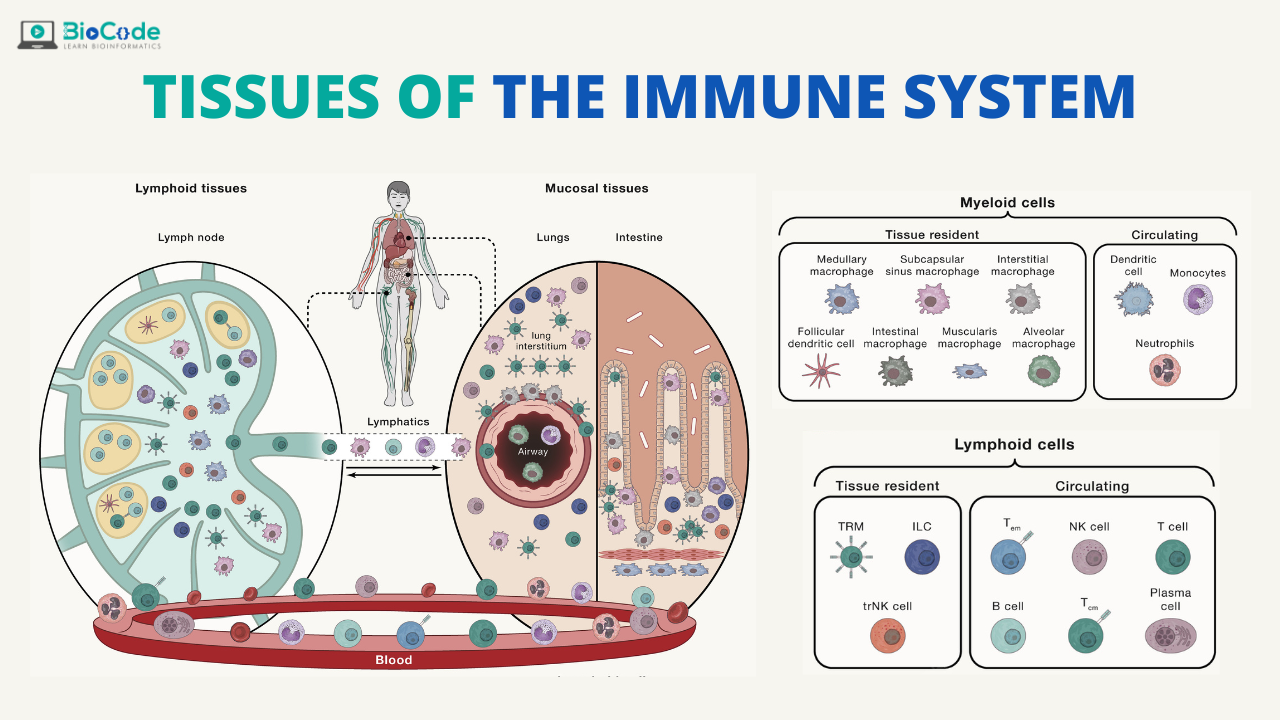
Cutaneous Immune System and Mucosal Immune System:
The cutaneous immune system and mucosal immune system are specialized collections of lymphoid tissues and APCs located in and under the epithelia of the skin and the gastrointestinal and respiratory tracts.
Many commensal bacteria are present in the lumen. The mucus-secreting epithelium provides an innate barrier to microbial invasion. Specialized epithelial cells, such as M cells, promote the transport of antigens from the lumen into underlying tissues. Cells in the lamina propria, including dendritic cells, T lymphocytes, and macrophages, provide innate and adaptive immune defense against invading microbes; some of these cells are organized into specialized structures, such as Peyer’s patches in the small intestine. Immunoglobulin A (IgA) is a type of antibody abundantly produced in mucosal tissues that is transported into the lumen, where it binds and neutralizes microbes.
Lymphocyte Recirculation and Migration into Tissues:
Naive lymphocytes constantly recirculate between the blood and peripheral lymphoid organs. There they may be activated by antigens to become effector cells. The effector lymphocytes migrate from lymphoid tissues to sites of infection, where microbes are eliminated. Migration of effector lymphocytes to sites of infection is most relevant for T cells, because effector T cells have to locate and eliminate microbes at these sites. Plasma cells do not need to migrate to sites of infection; instead, they secrete antibodies, and the antibodies enter the blood. The naive T cells enter lymph nodes through specialized postcapillary venules, called high endothelial venules (HEVs).
Migration of T Lymphocytes:
Naive T lymphocytes migrate from the blood through high endothelial venules into the T cell zones of lymph nodes, where the cells are activated by antigens. Activated T cells exit the nodes, enter the bloodstream, and migrate preferentially to peripheral tissues at sites of infection and inflammation.