The innate immune system has to distinguish microbes from host tissue within minutes, without the benefit of the gene rearrangement that gives lymphocytes their specificity. It does this with a limited set of germline-encoded receptors that recognise molecular patterns shared across whole classes of pathogen — structures that microbes cannot easily discard because they are essential to their survival.
These are the pattern recognition receptors (PRRs), and the structures they detect are pathogen-associated molecular patterns (PAMPs). The same receptors also detect damage-associated molecular patterns (DAMPs) released by injured or dying host cells, which is why sterile injury can produce inflammation that looks much like infection.
Why the innate system targets patterns rather than individual molecules
Adaptive immunity can generate receptors against essentially any structure, but it takes days to do so. Innate immunity must respond immediately, so it relies on a few hundred receptor specificities encoded directly in the genome.
This works because the targets are chosen carefully. Lipopolysaccharide, peptidoglycan, flagellin and double-stranded RNA are all either structural necessities or essential to replication. A bacterium cannot stop making peptidoglycan to evade detection without ceasing to be viable. The innate system exploits that constraint — and because these structures are absent from host cells, recognition is also a reliable test of foreignness.
Toll-like receptors
Toll-like receptors are the best characterised PRR family. Humans have ten functional TLRs, distributed between two locations, and that distribution is itself a recognition strategy.
Cell-surface TLRs: detecting microbial structure
- TLR4 — lipopolysaccharide from Gram-negative bacteria, with the accessory proteins MD2 and CD14. The central receptor in Gram-negative sepsis.
- TLR2, pairing with TLR1 or TLR6 — bacterial lipopeptides, peptidoglycan and lipoteichoic acid, mainly from Gram-positive organisms. Its ability to heterodimerise broadens the range of lipopeptides it can distinguish.
- TLR5 — flagellin, the protein subunit of the bacterial flagellum.
Endosomal TLRs: detecting microbial nucleic acid
- TLR3 — double-stranded RNA, produced during the replication of many viruses.
- TLR7 and TLR8 — single-stranded RNA.
- TLR9 — unmethylated CpG DNA, abundant in bacterial and viral genomes but rare and largely methylated in vertebrates.
Their location inside endosomes is the key to self–non-self discrimination. Nucleic acid is chemically similar whatever its source, so the system distinguishes by context: host DNA and RNA do not normally reach the endosomal compartment, whereas the contents of a phagocytosed microbe do. When this compartmentalisation fails, self nucleic acid activates these receptors — a mechanism implicated in systemic lupus erythematosus.
What happens after engagement
TLR ligation recruits adaptor proteins, principally MyD88 (used by all TLRs except TLR3) and TRIF (used by TLR3 and TLR4). MyD88 signalling activates NF-κB and the MAP kinases, driving inflammatory cytokines such as TNF, IL-1 and IL-6. TRIF signalling activates IRF3 and drives type I interferons, the core antiviral response.
That TLR4 can use both adaptors explains why LPS produces such a broad and powerful reaction — and why it can be so damaging when the response becomes systemic.
NOD-like receptors and the inflammasome
TLRs monitor the cell surface and endosomes. Cytosolic invaders require different sensors, and NOD-like receptors provide them.
NOD1 and NOD2 detect peptidoglycan fragments in the cytosol and signal through RIP2 to activate NF-κB. Loss-of-function NOD2 variants are among the strongest known genetic risk factors for Crohn’s disease, linking impaired bacterial sensing to chronic intestinal inflammation.
NLRP3 is the best known inflammasome-forming NLR. On activation it assembles with the adaptor ASC and pro-caspase-1 into a large complex that cleaves pro-IL-1β and pro-IL-18 into their active forms and triggers pyroptosis, an inflammatory form of cell death.
NLRP3 is unusual in that it does not appear to bind a specific ligand. Instead it responds to indicators of cellular disturbance — potassium efflux, lysosomal rupture, mitochondrial reactive oxygen species — which is why such varied stimuli converge on it, from bacterial pore-forming toxins to monosodium urate crystals in gout and cholesterol crystals in atherosclerosis.
RIG-I-like receptors: cytosolic viral RNA
Viruses replicating in the cytosol never enter the endosomal compartment, so endosomal TLRs cannot see them. RIG-I and MDA5 fill that gap.
RIG-I recognises short double-stranded RNA bearing a 5′-triphosphate — a signature of viral replication, since host cytosolic RNAs are capped or processed to remove it. MDA5 detects longer double-stranded RNA. Both signal through the mitochondrial adaptor MAVS to induce type I interferons.
Once again the discrimination is chemical rather than sequence-based: it is the modification state of the RNA end, not its sequence, that marks it as foreign.
C-type lectin receptors: recognising carbohydrate
C-type lectin receptors bind carbohydrate structures in a calcium-dependent manner and are especially important against fungi. Dectin-1 recognises β-glucans in fungal cell walls and drives antifungal immunity and Th17 responses; Dectin-2 and Mincle recognise mannose-containing structures and mycobacterial cord factor. The mannose receptor and DC-SIGN contribute to uptake and antigen presentation — and, in the case of DC-SIGN, are exploited by pathogens including HIV to gain entry to cells.
Scavenger receptors and complement receptors
Not every receptor’s job is to signal. Scavenger receptors such as SR-A and CD36 bind modified lipids and bacterial surface components and mediate phagocytosis, and their role in taking up oxidised LDL links them directly to foam cell formation in atherosclerosis.
Complement receptors recognise host-derived tags rather than microbial structures. When complement fragments such as C3b coat a microbe, CR1 and CR3 on phagocytes bind those fragments and greatly enhance uptake. This is opsonisation: the immune system labels a target, then reads its own label. Fc receptors work the same way for antibody-coated targets, forming one of the main bridges from adaptive immunity back to innate effector function.
Why the system uses several receptor families at once
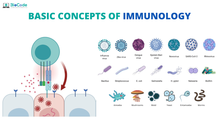
A single microbe engages many receptors simultaneously. A Gram-negative bacterium presents LPS to TLR4, flagellin to TLR5, peptidoglycan fragments to NOD1, and DNA to TLR9 once phagocytosed.
This redundancy serves two purposes. It is robust — losing one receptor does not blind the system. And it is informative: the particular combination of receptors engaged tells the cell what kind of threat it faces, and shapes the response accordingly. Predominantly TRIF-driven interferon signalling suits a virus; NF-κB-driven inflammatory cytokines suit an extracellular bacterium; Dectin-1 engagement biases towards the Th17 response that controls fungi.
Pattern recognition therefore does more than detect infection. It classifies it, and that classification determines which adaptive response is ultimately mounted.
Keep reading
Take it further
Learn this properly, with the work in front of you
Reading about it gets you the idea. Advanced Immunology MasterClass walks through the whole thing in order with videos you can follow at your own pace and a certificate at the end.
No free trial. Cancel whenever you want. Every course issues a verifiable certificate.


Join the discussionLeave a comment ↗